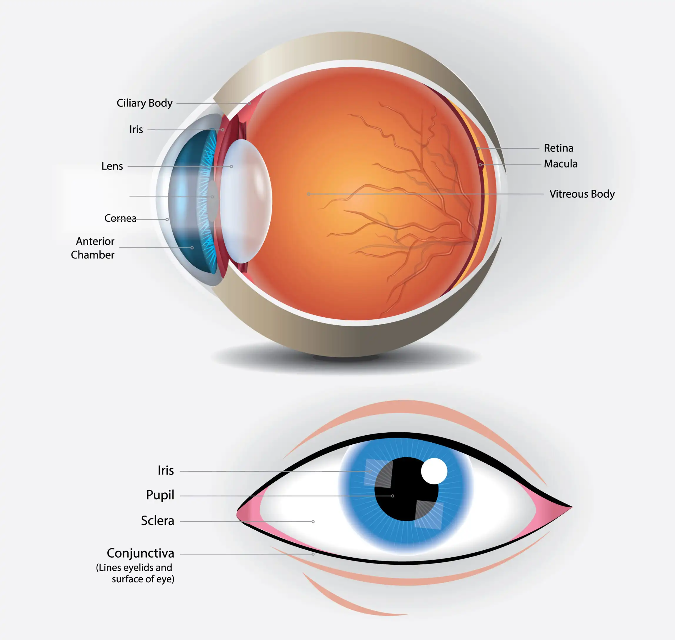
What is Cornea?
The outermost layer of the eye is called Cornea. It is one of the essential components of the human eye as it allows the light to enter into the eye for us to have vision. A cornea is generally 12mm in length and 11mm in height. The cornea also contributes towards around 70 percent of the focusing power of the eye. That is the main reason why it is imperative to take care of the corneas.
Types of Cornea
- Keratitis: Inflammation of the cornea, often caused by infections, injuries, or wearing contact lenses for extended periods.
- Corneal dystrophy:A group of genetic disorders characterized by the accumulation of abnormal material in the cornea, affecting its clarity and function.
- Corneal ulcers: corneal ulcers are open sores on the cornea usually resulting from infections, trauma, or inadequate tear production.
- Fuchs’ dystrophy: A progressive disease that affects the corneal endothelium (inner layer), leading to fluid buildup and cloudy vision.
- Corneal abrasions: scratches or injuries to the cornea’s surface. Often caused by foreign objects, contact lenses, and trauma.
- Corneal infections: bacterial, viral, and fungal infections that can lead to inflammation and damage to the cornea
Symptoms of Cornea
Corneal diseases display various symptoms affecting the transparent front part of the eye. Common signs include:
- Blurred or distorted vision
- Sensitivity to light
- Redness and eye pain
- Excessive tearing
- The sensation of a foreign body in the eye
- Watery or fluid discharge
- Gradual vision loss
- Inflammation and discomfort
- Decreased visual acuity (the ability to distinguish shapes and the details of objects at a given distance).
Immediate medical attention is important to prevent complications and preserve vision, as some conditions may progress rapidly.
Cause of Cornea
Corneal diseases can arise from various causes, including:
- Blurred or distorted vision
- Sensitivity to light
- Redness and eye pain
- Excessive tearing
- The sensation of a foreign body in the eye
- Watery or fluid discharge
- Gradual vision loss
- Inflammation and discomfort
- Decreased visual acuity (the ability to distinguish shapes and the details of objects at a given distance).
Immediate medical attention is important to prevent complications and preserve vision, as some conditions may progress rapidly.
Diagnosis of Corneal Disease
The diagnosis of corneal disorders typically involves a comprehensive eye examination conducted by a cornea specialist. The process of diagnosis involves: At first, the corneal specialist will start by gathering information about the patient's symptoms, medical history, and any relevant factors such as recent injuries, contact lens use, or systemic conditions.
Then, a standard eye chart is used to assess the patient's visual acuity, measuring how well they can see at various distances.
After that a slit-lamp microscope allows the eye care professional to examine the cornea and other structures of the eye in detail. This is essential for identifying irregularities, opacities, or signs of infection.
- Corneal Topography: This diagnostic test maps the curvature of the cornea, providing detailed information about its shape. It is particularly useful for detecting conditions like keratoconus.
- Pachymetry: Pachymetry measures the thickness of the cornea. Changes in thickness can be indicative of certain corneal conditions.
- Tear Film Assessment: Evaluation of tear production and the quality of the tear film is important, especially for conditions like dry eye syndrome that can affect the cornea.
- Intraocular Pressure Measurement: Tonometry is performed to measure intraocular pressure, as elevated pressure may be a sign of conditions like glaucoma or other corneal issues.
- Fluorescein Staining: Fluorescein dye may be used to highlight irregularities on the corneal surface, such as abrasions or ulcers.
- Corneal Biopsy or Scraping (if needed): In cases of suspected infection, a sample of corneal tissue may be collected for laboratory analysis.
- Specialized Tests: Depending on the suspected condition, additional tests such as endothelial cell counts, confocal microscopy, or genetic testing may be performed.
Combining these diagnostic tools allows eye or cornea specialists to identify and characterize corneal diseases accurately. Timely and accurate diagnosis is essential for developing an appropriate treatment plan and managing corneal conditions effectively.
Corneal Transplantation
Corneal transplantation, also known as corneal grafting, is a surgical procedure in which a damaged or diseased cornea is replaced with a healthy cornea from a donor. This procedure is often performed to restore vision, relieve pain, and improve the appearance of the eye. There are different types of corneal transplantation procedures, and the choice depends on the specific condition and the layers of the cornea affected.
The two main types are penetrating keratoplasty (PK) and endothelial keratoplasty.
Penetrating Keratoplasty (PK)
- Procedure: IIn a PK, the entire thickness of the central cornea is removed and replaced with a donor cornea. The surgeon uses a circular trephine to remove a button-shaped section of the patient's cornea, and the donor cornea is then sutured in place.
- Indications: PK is used for conditions that affect the full thickness of the cornea, such as advanced keratoconus, corneal scarring, or corneal dystrophies.
Endothelial Keratoplasty (EK)
- Descemet's Stripping Endothelial Keratoplasty (DSEK): This procedure involves replacing the innermost layers of the cornea, including the endothelium and Descemet's membrane, with a donor graft.
- Descemet's Membrane Endothelial Keratoplasty (DMEK): Similar to DSEK, It involves replacing the Descemet membrane and endothelium with donor tissue:
EK procedures are commonly used for conditions affecting the endothelium, such as Fuchs' endothelial dystrophy or corneal edema.
Procedure: The surgical procedure is typically performed under local or general anesthesia.
The damaged cornea is carefully removed, and the donor cornea is sized and sutured or adhered in place.
Sutures may be used to secure the graft initially, and they may be removed or remain in place depending on the specific case.
Following surgery, patients undergo a period of recovery and are closely monitored for signs of rejection or other complications.
Recovery and Follow-Up: The recovery period varies, but patients may experience gradual improvement in vision over several months.
Regular follow-up visits with the best cornea specialist are essential to monitor the healing process and address potential issues.
Corneal Transplantation is a highly successful procedure, and the outcome depends on various factors, including the patient's overall eye health and the specific condition being treated. As with any surgery, there are risks and potential complications, and individuals considering corneal transplantation should discuss these with their eye care professional.
The choice of treatment depends on the severity of the disease, the patient's lifestyle and preferences, and the response to earlier interventions.
Keratoconus (Corneal Disease)
Keratoconus is a progressive eye disorder that causes the cornea to thin and takes on a cone-like shape, resulting in distorted vision. Generally, this condition begins between the ages of 10 to 25. keratoconus Treatment aims to improve visual function, manage symptoms, and in some cases, slow the progression of the condition.
Treatment options for keratoconus
- Contact lenses and eyeglasses: In the initial stage of keratoconus contact lenses or eyeglasses may be sufficient.
- Rigid Gas Permeable (RGP) Contact Lenses: RGP lenses are often prescribed for moderate to advanced keratoconus. These lenses provide a smooth, rigid surface that helps to correct the irregular shape of the cornea, improving vision.
- Hybrid Contact Lenses: Hybrid lenses have a rigid center surrounded by a soft outer ring. They aim to provide the comfort of soft lenses with the visual clarity of RGP lenses.
- Scleral Contact Lenses: These large-diameter lenses vault over the cornea, creating a tear-filled space between the lens and the cornea. Scleral lenses can provide better comfort for some patients with advanced keratoconus.
- Corneal Cross-Linking (CXL): Corneal cross-linking is a procedure designed to strengthen the cornea and prevent further progression of keratoconus. It involves applying riboflavin (vitamin B2) eye drops to the cornea, followed by exposure to ultraviolet (UV) light. This strengthens the corneal collagen fibers.
- Intacs (Intrastromal Corneal Rings): Intacs are small, crescent-shaped devices that are surgically implanted into the cornea. They help flatten the cornea, improving its shape and vision.
- Phakic Intraocular Lenses (IOLs): In some cases, particularly when contact lenses are not well-tolerated, phakic intraocular lenses may be considered. These are implanted without removing the natural lens and can correct refractive errors associated with keratoconus.
- Corneal Transplant (Penetrating Keratoplasty or Descemet's Stripping Endothelial Keratoplasty): In advanced cases where other treatments are not effective, a corneal transplant may be recommended. This involves replacing the damaged corneal tissue with healthy donor tissue.