Cataract Treatment
What Is Cataract?
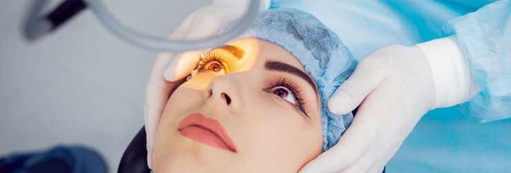
C-lasik corrects the spectacle power along optical aberrations in an eye resulting in a sharp vision. Optical aberrations are measured with a machine called aberrometer.
Surgical Technique
Cataracts are changes in clarity of the natural lens inside the eye that gradually degrade visual quality. The natural lens sits behind the colored part of the eye (iris) in the area of the pupil, and cannot be directly seen with the naked eye unless it becomes extremely cloudy. The lens plays a crucial role in focusing unimpeded light on the retina at the back of the eye. The retina transforms light to a neurologic signal that the brain interprets as vision. Significant cataracts block and distort light passing through the lens, causing visual symptoms and complaints.
The term cataract is derived from the Greek word cataractos, which describes rapidly running water. When water is turbulent, it is transformed from a clear medium to white and cloudy. Keen Greek observers noticed similar-appearing changes in the eye and attributed visual loss from "cataracts" as an accumulation of this turbulent fluid, having no knowledge of the anatomy of the eye or the status or importance of the lens.
CAUSES OF CATARACT
The lens is made mostly of water and protein. Specific proteins within the lens are responsible for maintaining its clarity. Over many years, the structures of these lens proteins are altered, ultimately leading to a gradual clouding of the lens. Rarely, cataracts can present at birth or in early childhood as a result of hereditary enzyme defects, and severe trauma to the eye, eye surgery, or intraocular inflammation can also cause cataracts to occur earlier in life. Other factors that may lead to development of cataracts at an earlier age include excessive ultraviolet-light exposure, diabetes, smoking, or the use of certain medications, such as oral, topical, or inhaled steroids. Other medications that are more weakly associated with cataracts include the long-term use of statins and phenothiazines
Cataract Symptoms
Having cataracts is often compared to looking through a foggy windshield of a car or through the dirty lens of a camera. Cataracts may cause a variety of complaints and visual changes, including blurred vision, difficulty with glare (often with bright sun or automobile headlights while driving at night), dulled color vision, increased nearsightedness accompanied by frequent changes in eyeglass prescription, and occasionally double vision in one eye. Some people notice a phenomenon called "second sight" in which one's reading vision improves as a result of their increased nearsightedness from swelling of the cataract
Surgery
The standard cataract surgical procedure is typically performed in either a hospital or in an ambulatory surgery center. The most common form of cataract surgery today is a process called phacoemulsification. With the use of an operating microscope, your surgeon will make a very small incision in the surface of the eye in or near the cornea. A thin ultrasound probe is inserted into the eye that uses ultrasonic vibrations to dissolve (phacoemulsify) the clouded lens. These tiny fragmented pieces are then suctioned out through the same ultrasound probe. Once the cataract is removed, an artificial lens is placed into the same thin capsular bag that the cataract occupied. This intraocular lens is essential to help your eye focus after surgery.
There are three basic techniques for cataract surgery:
- Phacoemulsification:
This is the most common form of cataract removal as explained above. In this most modern method, cataract surgery can usually be performed in less than 30 minutes and usually requires only minimal sedation and numbing drops, no stitches to close the wound, and no eye patch after surgery.
- Extracapsular cataract surgery:
This procedure is used mainly for very advanced cataracts where the lens is too dense to dissolve into fragments (phacoemulsify) or in facilities that do not have phacoemulsification technology. This technique requires a larger incision so that the cataract can be removed in one piece without being fragmented inside the eye. An artificial lens is placed in the same capsular bag as with the phacoemulsification technique. This surgical technique requires a various number of sutures to close the larger wound, and visual recovery is often slower. Extracapsular cataract extraction usually requires an injection of numbing medication around the eye and an eye patch after surgery.
- Intracapsular cataract surgery:
This surgical technique requires an even larger wound than extracapsular surgery, and the surgeon removes the entire lens and the surrounding capsule together. This technique requires the intraocular lens to be placed in a different location, in front of the iris. This method is rarely used today but can be still be useful in cases of significant trauma
As the natural lens plays a vital role in focusing light for clear vision, artificial-lens implantation at the time of cataract surgery is necessary to yield the best visual results. Because the implant is placed in or near the original position of the removed natural lens, vision can be restored, and peripheral vision, depth perception, and image size should not be affected. Artificial lenses are intended to remain permanently in place, require no maintenance or handling, and are neither felt by the patient nor noticed by others.
There are a variety of intraocular lens styles available for implantation, including monofocal, toric, and multifocal intraocular lenses.
- Monofocal lens:
These lenses are the most commonly implanted lenses today. They have equal power in all regions of the lens and can provide high-quality vision at a single focal point (usually at distance). They usually require only a light pair of spectacles for optimal distance vision correction. However, monofocal lenses do not correct astigmatism, an irregular oblong corneal shape that can distort vision at all distances, and require corrective lenses for all near tasks, such as reading or writing.
- Toric lens:
Toric lenses have more power in one specific region in the lens (similar to spectacles with astigmatism correction in them) to correct astigmatism, which can further improve unaided distance vision for many individuals. Due to the difference in lens power in different areas, the correction of astigmatism with a toric lens requires that the lens be positioned in a very specific configuration. While toric lenses can improve distance vision and astigmatism, they still require corrective lenses for all near tasks, such as reading or writing.
- Multifocal lens:
Multifocal intraocular lenses have a variety of regions with different power within the lens that allows individuals to see at a variety of distances, including distance, intermediate, and near. While promising, multifocal lenses are not for everyone. They can cause significantly more glare than monofocal or toric lenses. Further, multifocal lenses cannot correct astigmatism, and some patients require additional surgery such as LASIK to correct astigmatism and maximize their unaided vision.